
ECG test results explained in simple words—finally understand what your ECG report really means for your heart.
Most ECG changes are not dangerous—many are mild, temporary, or completely harmless.
A patient-friendly guide by RealMedVision
Last Updated — May 2026

You Got Your ECG Report — Now What?
Many people panic after seeing an abnormal ECG report. The lines look strange, the words feel technical, and nobody really explains what any of it means. That is exactly why ECG test results explained in simple words matter so much for patients and families.
You sit there staring at terms like “ST depression” or “T wave inversion,” and your mind immediately jumps to the worst possible conclusion.
Here is the truth: getting your ECG test results explained in simple words can remove a lot of unnecessary fear and confusion. Most ECG changes are not dangerous. Many are caused by stress, anxiety, caffeine, or simply how your body was feeling that day. Doctors never judge an ECG alone.
They always look at the full picture—your symptoms, your history, and how you are feeling right now.
This article gives you exactly that—ECG test results explained in simple words, without scary medical language, without confusing charts, and without making you feel like you are reading a textbook.
What This Article Covers
- What ECG test results actually mean
- Normal ECG results explained simply
- Abnormal ECG signs explained in simple words
- Can ECG detect heart attack or blockage
- Symptoms that need immediate attention
- What causes abnormal ECG results
- Can stress or anxiety affect your ECG
- ECG Test vs 2D Echo—what is the difference
- When should you actually worry
- Lifestyle tips for better heart health
- FAQs answered simply
ECG Test Results Explained in Simple Words: What They Mean
An ECG, Electrocardiogram—records the electrical activity of your heart. Every single heartbeat produces a tiny electrical signal. The ECG machine captures these signals and draws them as waves and lines on paper or a screen.
These waves are not random. Each one has a name and a specific meaning. When doctors read your ECG report, they are looking at how these waves are shaped, how fast they appear, and whether the gaps between them are normal.
Think of it like this—imagine your heart is speaking in electrical pulses. The ECG is simply a recording of that conversation.
ECG test results explained in simple words become much easier to understand when you see the ECG as your heart’s electrical voice. When the conversation flows smoothly and regularly, everything is fine. When something interrupts that flow, the ECG picks it up.
Getting your ECG test results explained in simple words means understanding what each part of that conversation is saying, and that is exactly what this article is here to do.
Normal ECG Results Explained in Simple Words
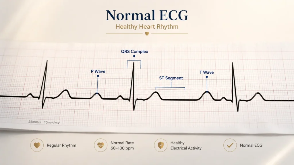
A normal ECG means your heart’s electrical system was working properly at the time of the test. This sounds simple and it actually is.
Here is what a normal ECG looks like, explained without the medical jargon:
Heart Rate —
Heart Rate: Between 60 and 100 beats per minute at rest. This is the healthy range confirmed by the American Heart Association.
Heart Rhythm —
Heart Rhythm: Regular and consistent. Each beat follows the last one at an even pace, like a steady clock ticking.
P Wave —
P Wave: A small gentle bump before the main spike. It means the upper chambers of the heart—called the atria—are contracting normally and pushing blood into the lower chambers.
QRS Complex —
QRS Complex: The tall sharp spike you see on every ECG. This is the most important wave. It means the lower chambers—the ventricles—are pumping blood out to your lungs and body. A normal QRS is narrow and sharp.
T Wave —
T Wave: The gentle wave that follows the big spike. It shows the heart muscle recovering and resetting for the next beat. Normal T waves are smooth and upright.
ST Segment —
ST Segment: The flat line between the QRS spike and the T wave. In a healthy heart, this sits exactly at the baseline—not elevated, not depressed.
Many people also hear about Ejection Fraction: this is how much blood the heart pumps with each beat. It is not measured by ECG alone but is usually assessed through a 2D Echo test. Both tests together give doctors a much more complete picture of heart health.
Many healthy people also show mild ECG changes that are completely harmless. ECG test results explained in simple words often surprise people because small ECG variations can happen due to age, body type, fitness level, stress, and even normal breathing patterns.
Abnormal ECG Signs Explained in Simple Words
This is the section most people are actually looking for. This sounds scary, but please read it fully before worrying.
Tachycardia —
Heart rate above 100 beats per minute at rest. Your heart is beating too fast. This commonly happens with stress, fever, dehydration, thyroid problems, too much caffeine, or anxiety. It is not always a sign of heart disease.
Bradycardia —
Heart rate below 60 beats per minute. The heart is beating slowly. In athletes and very fit people this is completely normal. In others it may need a doctor’s evaluation.
Irregular Heartbeat (Arrhythmia) —
The beats are not evenly spaced. Sometimes the heart skips a beat or adds an extra one. Many people live with occasional skipped beats and never even notice them. Conditions like Atrial Fibrillation are more serious and need proper management.
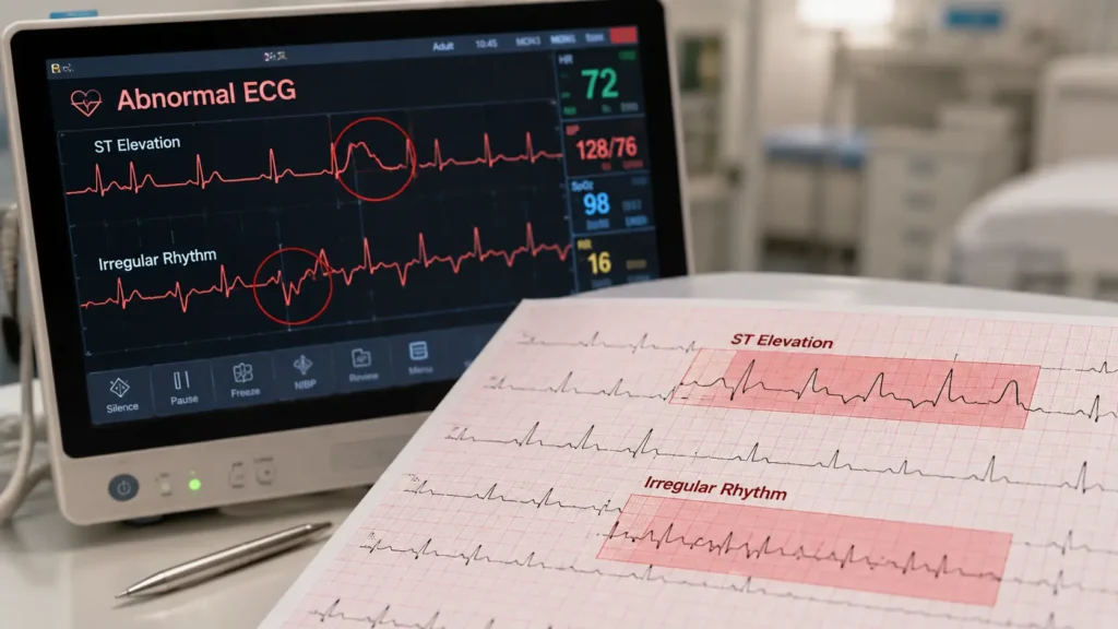
ST Elevation —
The ST segment rises above the baseline. In the presence of chest pain, this is one of the most urgent ECG findings. It strongly suggests a STEMI—ST Elevation Myocardial Infarction, a serious heart attack where a coronary artery is completely blocked. According to Mayo Clinic, STEMI requires immediate emergency treatment.
ST Depression —
The ST segment drops below the baseline. This may suggest NSTEMI—a less severe type of heart attack—or Angina, which means reduced blood flow to the heart. High cholesterol and blocked arteries are common causes behind these changes.
T Wave Inversion —
The T wave flips downward instead of pointing up. This can suggest reduced blood supply to the heart muscle, called ischemia. This sounds scary, but it is not always dangerous. Many causes are temporary and treatable.
Heart Block —
Electrical signals are delayed between the upper and lower chambers. First degree heart block is mild and often needs no treatment. Third degree is serious and needs urgent attention.
Doctors never judge an ECG alone. ECG test results explained in simple words become more accurate when doctors compare the ECG pattern with your symptoms, medical history, and overall heart health.
As explained in Dale Dubin’s Rapid Interpretation of EKGs, ECG findings should always be interpreted in clinical context for the correct diagnosis.
Quick Summary: Normal vs Abnormal ECG Results
ECG Finding | What It May Mean |
|---|---|
Normal Rhythm | Healthy heart electrical activity |
ST Elevation | Possible STEMI—emergency |
ST Depression | Angina or NSTEMI |
Fast Heartbeat (100+ BPM) | Stress, anxiety, or Tachycardia |
Slow Heartbeat (below 60 BPM) | Bradycardia |
T-wave inversion | Possible ischemia |
Irregular Rhythm | Possible Arrhythmia or AFib |
Heart Block Pattern | Electrical conduction delay |
Can ECG Detect Heart Attack or Blockage?
Yes, but with important limits every patient should know.
During an active heart attack, the ECG almost always shows clear changes. STEMI—complete blockage—shows up as ST elevation and is diagnosed within minutes. NSTEMI and Angina—partial blockage or reduced blood flow may show ST depression or T wave changes.
This is why hospitals perform an ECG within minutes of any chest pain presentation. Every minute of delay means more heart muscle at risk.
However, a resting ECG can be completely normal even when significant blocked arteries are present.
The narrowed artery may only cause problems when the heart is under physical stress. In this situation, doctors recommend a stress test, Holter monitor, or coronary angiography to get the full picture.
A normal ECG does not guarantee a perfectly healthy heart. ECG test results explained in simple words can still look normal even when some heart problems are developing silently. It simply means the electrical activity looked normal at the moment of recording.
If symptoms continue despite a normal ECG, always follow up with your doctor.
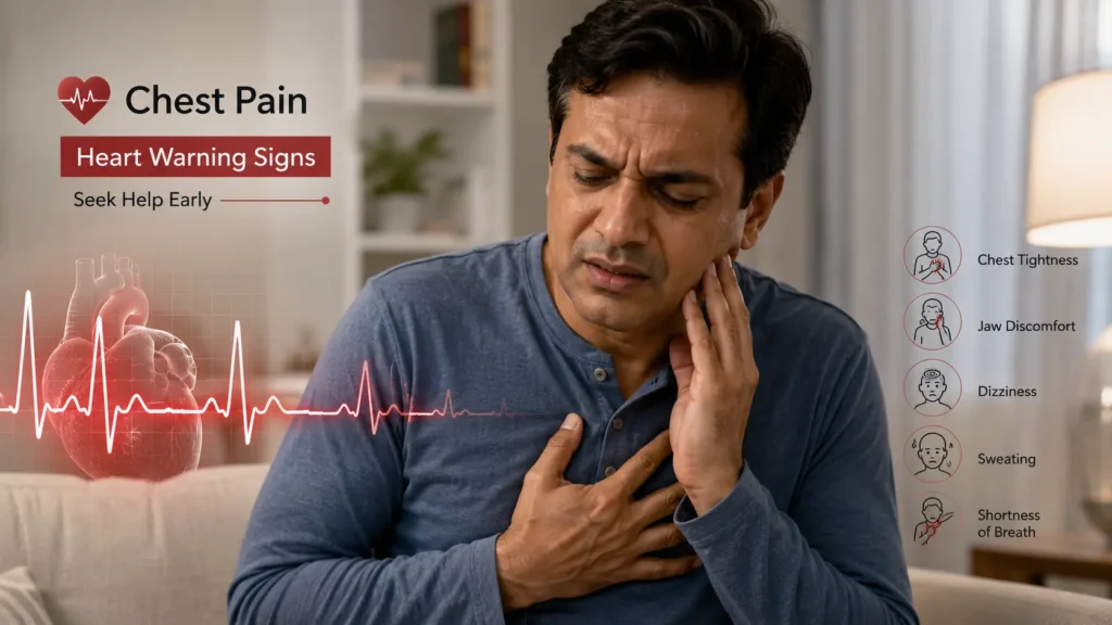
Symptoms That Need Immediate Attention
Symptom | ECG Needed? |
|---|---|
Chest pain or tightness | Yes—immediately |
Fast or racing heartbeat | Often |
Fainting or blackout | Yes |
Breathlessness at rest | Often |
Dizziness or lightheadedness | Often |
Irregular heartbeat feeling | Yes |
Pain spreading to arm or jaw | Yes—emergency |
If chest pain comes with breathlessness, sweating, or fainting, do not wait. Call emergency services immediately.
What Causes Abnormal ECG Results?
Not every abnormal ECG means heart disease. Many completely healthy people show minor changes because of everyday lifestyle factors.
Lifestyle causes: stress, anxiety, too much caffeine, smoking, alcohol, poor sleep, and dehydration can all temporarily change your ECG pattern. These are very common and very reversible.
Medical causes: thyroid problems; electrolyte imbalance such as low potassium or magnesium,
and certain medicines including blood pressure drugs and antidepressants can affect ECG readings.
Heart conditions: Rheumatic Heart Disease, Congenital Heart Disease present from birth, Peripheral Artery Disease, and Aortic Regurgitation all produce specific abnormal ECG patterns that doctors are trained to recognize.
According to WHO, cardiovascular diseases cause approximately 17.9 million deaths every year globally. ECG test results explained in simple words can help people recognize early warning signs and understand when medical attention may be needed.
Can Stress or Anxiety Affect Your ECG?
Absolutely yes—and this is far more common than most people realize.
When you feel anxious, your body releases adrenaline. This speeds up your heart rate, which may appear on the ECG as tachycardia. ECG test results explained in simple words can sometimes look frightening, even when the changes are caused by stress or anxiety rather than actual heart disease.
Minor ST and T wave changes may look abnormal on paper but are often temporary and harmless.
Many patients come in with ECG changes that completely normalize once they calm down and the test is repeated. This is not unusual at all.
Doctors also look at stress, sleep, caffeine intake, and anxiety before reading an ECG. ECG test results explained in simple words always make more sense when doctors understand the full situation.
ECG Test vs 2D Echo: What Is the Difference?
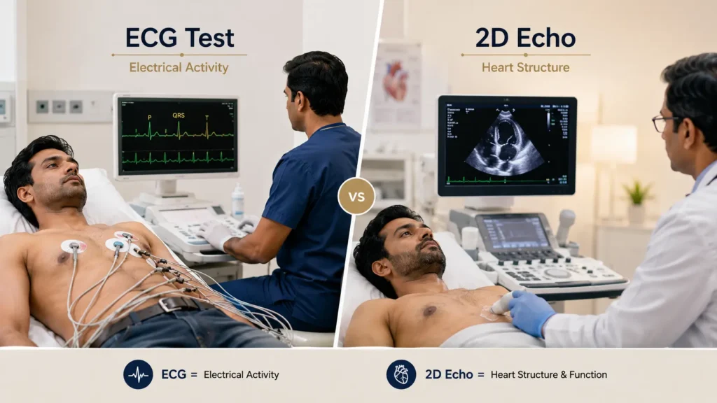
This is a question many patients ask—and it is a very good one.
ECG records the electrical activity of the heart. It tells you how fast the heart is beating, whether the rhythm is regular, and whether the electrical pathways are working correctly.
It takes about 5 to 10 minutes and is completely painless.
2D Echo (Echocardiogram) uses sound waves—like an ultrasound—to create a moving image of the heart. It shows the physical structure of the heart, how the valves are working, and the Ejection Fraction, how strongly the heart is pumping blood with each beat.
Simply put, ECG checks the heart’s electrical system. 2D Echo checks the heart’s physical structure and pumping strength.
Both tests measure different things. Doctors often order both together to get a complete picture. If your ECG shows something unusual, a 2D Echo is usually the next step to understand what is actually happening structurally inside the heart.
When Should You Worry About ECG Results?
Do not panic if the report shows mild tachycardia, occasional skipped beats, or small T wave changes without any symptoms. These are often harmless and related to lifestyle factors. Many healthy people show exactly these findings.
See your doctor soon if the report mentions bundle branch block, prolonged QT interval, or persistent arrhythmia—even without symptoms. These need evaluation but are rarely emergencies.
Seek emergency help immediately if you have chest pain, breathlessness, or fainting along with ST elevation or severe rhythm changes. This combination always needs urgent attention.
The NIH and American Heart Association both emphasize that early evaluation of ECG changes alongside symptoms leads to significantly better outcomes for patients.
Lifestyle Tips for Better Heart Health
Small daily habits make a real and measurable difference to your heart over time.
Control cholesterol —
High cholesterol builds plaque in arteries slowly over years, eventually causing blocked arteries and the ST changes we discussed earlier. A diet rich in vegetables, whole grains, and healthy fats helps keep cholesterol in check.
Maintain normal blood pressure —
Sustained high blood pressure thickens the heart muscle, and this shows up on ECG over time. Regular monitoring and lifestyle management matter enormously.
Exercise regularly —
Even 30 minutes of brisk walking five days a week strengthens the heart muscle and improves its electrical efficiency. Your ECG actually looks healthier with regular exercise.
Quit smoking —
Smoking damages blood vessel walls and accelerates plaque buildup. The connection between smoking and heart attack risk is direct and well proven.
Manage stress —
Chronic stress keeps adrenaline elevated, which over time affects both heart rhythm and blood pressure. Simple daily practices like deep breathing, walking, or good sleep go a long way.
Sleep well —
Poor sleep is directly linked to irregular heartbeat and higher cardiovascular risk according to recent WHO data. Seven to eight hours of consistent sleep is genuinely good for your heart.
Frequently Asked Questions
What do ECG test results explained in simple words actually mean?
It means understanding your ECG report without medical jargon — knowing whether your heart rate, rhythm, and wave patterns are normal, and what any abnormal findings actually indicate in everyday language.
Can an ECG detect blocked arteries?
It can suggest blockage when ST or T wave changes are present—especially if symptoms like chest pain are also there. But a resting ECG cannot confirm blocked arteries on its own. A stress test or angiography is needed for that confirmation.
Can anxiety cause an abnormal ECG?
Yes. Anxiety raises adrenaline which speeds up the heart rate and can cause minor wave changes. These typically normalise once the anxiety passes and are not caused by actual heart disease.
Can an ECG detect weak heart muscles?
Partially. An ECG can show certain patterns that suggest heart muscle damage, like abnormal Q waves after a previous heart attack. But assessing actual pumping strength requires a 2D Echo and Ejection Fraction measurement.
Is ECG enough to detect all heart disease?
No. ECG is a powerful first step but it has limitations. It captures electrical activity at one moment in time. Structural problems, valve disease, and intermittent arrhythmias may need 2D Echo, Holter monitoring, or angiography to properly diagnose.
What happens after an abnormal ECG?
Your doctor will review your symptoms and history alongside the report. They may order additional tests—2D Echo, stress test, blood tests, or Holter monitor—depending on what the ECG shows and how you are feeling clinically.
Is an abnormal ECG always dangerous?
Not at all. Many abnormal findings are mild, harmless, or caused by temporary factors like stress or caffeine. Always have your result reviewed by a doctor but do not assume the worst before speaking to one.
What is the normal ECG range?
Normal heart rate is 60 to 100 beats per minute. A normal PR interval is 120 to 200 milliseconds. Normal QRS duration is less than 120 milliseconds. The ST segment should sit exactly at the baseline.
Can cholesterol affect ECG results?
Not directly. But high cholesterol leads to blocked arteries over time, which then causes ST and T wave changes on the ECG during ischemia or a heart attack.
Conclusion
If there is one thing to take away from this article, it is this. An ECG is a tool, not a verdict.
Getting your ECG test results explained in simple words is not about memorizing wave names or medical terms. It is about understanding that your heart gave doctors important information—and that information, in the right hands, leads to better care and better outcomes.
If your report showed something unexpected, do not panic. Talk to your doctor. Ask what it means. Understand what the next step is. Early awareness combined with honest lifestyle changes and timely medical review—these three things together make the biggest difference to your long-term heart health.
Your heart is worth that attention. Always has been.
Medical Disclaimer
This article is written for general educational awareness only. It does not constitute medical advice, diagnosis, or treatment recommendations. If you are experiencing heart-related symptoms, please consult a qualified doctor promptly. Do not delay medical evaluation based on information in this article. References include WHO, NIH, American Heart Association, Mayo Clinic, and Dale Dubin’s Rapid Interpretation of EKGs.
About the Author
Iraphan Khan, BSN | D.Pharm | CMLT, is a Healthcare SEO Strategist and Medical Content Writer at RealMedVision, creating clinically accurate content optimized for Google and AI search.
Medically Reviewed By
Dr Praveen Verma, MBBS, MD — Diagnostic & Pathology
Dr Himanshu Morya MBBS — Clinical Accuracy & Patient Safety
Kalpna Singh Shekhawat BSN NP — Patient Care & Practical Accuracy
References:
1. American Heart Association — ECG Guidelines
2. Mayo Clinic — Electrocardiogram Overview
3. NIH — National Heart, Lung, and Blood Institute
4. WHO — Cardiovascular Disease Statistics
5. MedlinePlus — ECG Test Information
6. Cleveland Clinic — ECG Results Explained
7. Harvard Health — Understanding Your ECG
8. British Heart Foundation — ECG Explained
9. NHS UK — Electrocardiogram ECG
10. Dale Dubin — Rapid Interpretation of EKGs
11. American College of Cardiology — ECG Abnormalities
12. PubMed — ECG Clinical Studies