
You just got your 2D Echo. You see EF 60%—and you want to know: is this dangerous?
EF 60% means your heart is pumping well. That is good news.
A patient-friendly guide by RealMedVision
Last Updated — May 2026

You just got your 2D Echo report. You see one number—EF 60%. And instantly the fear starts. Is this dangerous? Is something wrong with my heart?
Most patients panic after seeing EF 60% in 2D Echo report—not because something is wrong, but because nobody explained what this number actually means.
This guide explains it simply. The way a cardiologist explains it to patients sitting right across the table.
What Is EF in a 2D Echo Report?
EF stands for Ejection Fraction. It is a percentage that tells you how much blood your heart pushes out with every single heartbeat.
Your heart is a pump. It fills with blood and pushes that blood to the rest of your body with every heartbeat. But the heart does not empty completely each time it beats. The percentage of blood pushed out is called the Ejection Fraction, or EF.
In a 2D Echo report, EF helps doctors understand how well your heart is pumping.
Here is a simple way to understand it. Your left ventricle—the main pumping chamber—fills with roughly 100 ml of blood before each beat. If it pushes out 60 ml and keeps 40 ml behind, your EF is 60%. That leftover blood is not a problem. It is completely normal physiology.
This number is measured during a 2D Echo test, which uses sound waves to create real-time images of your heart. It is painless, completely safe, involves no radiation, and takes about 15 to 30 minutes.
Is EF 60% Normal or Dangerous?
Let me answer this directly—EF 60% in 2D Echo report is completely normal. It is not dangerous.
According to the American Heart Association, a normal ejection fraction usually falls between 55% and 70%. So if your report shows EF 60% in a 2D Echo, it generally means your heart is pumping blood normally and efficiently.
In most cases, EF 60% is deeply reassuring. It means your heart muscle is strong, your left ventricle is contracting well, and there is no pumping failure.
Many patients ask, if EF 60% in a 2D Echo is normal, why is the heart not pumping 100%? The answer is simple: the heart is not designed to empty completely with every beat. Keeping some blood inside the heart is completely normal. In most cases, EF 60% in a 2D Echo report means healthy heart function.
So if your report says EF 60%, take a breath. Your heart is doing exactly what it is supposed to do.
What Is a Normal Ejection Fraction Range?

Understanding the full EF range helps you read any future report with confidence.
According to the American College of Cardiology and research published on NIH and Mayo Clinic, the classification is:
55% to 70% — Normal EF in 2D Echo. Your heart is pumping strongly. EF 60% sits right here.
50% to 54% — Low Normal. Slightly below midpoint but acceptable in most patients without symptoms.
41% to 49% — Mildly Reduced EF. Pumping slightly below normal. A cardiologist will monitor carefully. Symptoms may appear during physical activity.
40% or below — Reduced EF. This is where heart failure with reduced ejection fraction begins. Medical treatment is needed.
Below 30% — Severely Reduced. Serious cardiac dysfunction requiring immediate management.
Is EF 60 normal? Absolutely yes—it falls well within the normal range
How to Read EF in Your 2D Echo Report
When you receive your 2D Echo report, most patients do not know where to look first. Here is a simple, practical guide.
Find the line that says “EF” or “Ejection Fraction” — it is usually expressed as a percentage. If the number is between 55% and 70%, that section of your report is normal.
Next, look for “LV Function” — if it says “normal LV function” or “good LV systolic function,” that is reassuring. If it says “mildly impaired” or “reduced,” discuss with your cardiologist.
Then check the valve comments. Words like “mild MR” or “mild TR” mean very minor leakage—these are common findings in the general population and usually not serious. “Moderate” or “severe” next to any valve finding needs follow-up.
Finally, check whether “pericardial effusion” is mentioned. If the report says “no pericardial effusion,” that is normal and good.
Remember — EF 60% in 2D Echo report combined with normal LV function and no significant valve disease is an excellent result overall.
Can EF 60% Still Cause Symptoms?
This is a question many patients have but rarely ask—and it is an important one.
Yes, it is possible to have normal EF in 2D Echo and still experience symptoms like mild breathlessness, chest discomfort, or fatigue. This happens because EF measures only the pumping function of the left ventricle. It does not measure everything about your heart’s health.
Some patients may still have symptoms even when EF 60% in a 2D Echo report is normal. This can happen in conditions like diastolic dysfunction, where the heart becomes stiff and does not fill with blood properly.
Mild valve disease, high blood pressure, or narrowed coronary arteries can also cause symptoms even when EF appears normal.
According to Mayo Clinic, chest pain or shortness of breath during activity can happen even when EF 60% in a 2D Echo report appears normal. That is why doctors do not rely on just one number. They also look at your symptoms, blood pressure, cholesterol levels, and full clinical history together.
So if you have EF 60% in 2D Echo but still feel breathless or uncomfortable — do not dismiss those symptoms. Report them to your doctor. EF alone does not tell the complete story.
What Happens If EF Becomes Low?

When EF drops below normal, the body begins showing warning signs. Knowing these symptoms could save your life.
When the heart starts struggling, breathlessness hits first. Climbing one floor feels exhausting. Lying flat at night becomes uncomfortable. Legs and ankles start swelling.
Even if your EF 60% in 2D Echo report was normal before — new symptoms should never be ignored.
Fast or irregular heartbeats—called palpitations—are another warning. Dizziness, lightheadedness, and reduced ability to do normal daily activities all appear as EF falls further.
When EF drops below 30%, the risk of life-threatening complications—including sudden cardiac arrest—increases significantly. This is why routine monitoring and early detection matter so much.
Common Causes of Abnormal EF
EF does not drop randomly. Several well-known conditions damage the heart muscle over time.
Heart Attack
Heart attacks—both STEMI and NSTEMI types—are leading causes. When part of the heart muscle dies due to a blocked artery, that area loses its ability to contract, and EF falls. Heart attack symptoms like severe chest pain, sweating, and breathlessness should never be ignored.
High Blood Pressure
High Blood Pressure that stays uncontrolled for years forces the heart to work harder than it should. Eventually the muscle thickens and then weakens. Keeping normal blood pressure is one of the most powerful things you can do to protect your EF.
High Cholesterol
High Cholesterol builds plaque inside coronary arteries. This leads to angina first—chest tightness during exertion—and eventually to a heart attack if not treated with medicines and lifestyle changes.
Rheumatic Heart Disease
Rheumatic Heart Disease, which remains common in India, damages the mitral and aortic valves. Damaged valves force the heart to work inefficiently, gradually reducing EF over years.
Aortic Regurgitation
Aortic regurgitation, where the aortic valve leaks blood backwards into the left ventricle—places chronic volume overload on the heart. Over time this weakens the muscle.
Congenital Heart Disease
Congenital Heart Disease—structural defects present from birth—can affect EF depending on severity. Regular cardiology follow-up is essential in such cases.
Peripheral Artery Disease
Peripheral Artery Disease and other systemic conditions can also place indirect stress on cardiac function over years.
These two tests confuse patients constantly. They measure completely different things.
Feature | ECG Test | 2D Echo |
|---|---|---|
What it measures | Electrical activity | Structure and pumping function |
Shows EF? | No | Yes |
Best for | Rhythm, heart attack detection | EF, valve disease, chamber size |
Duration | 5–10 minutes | 15–30 minutes |
Radiation | None | None |
In most cardiac evaluations, doctors order both because each answers questions the other cannot.
ECG Test vs 2D Echo: What’s the Difference?
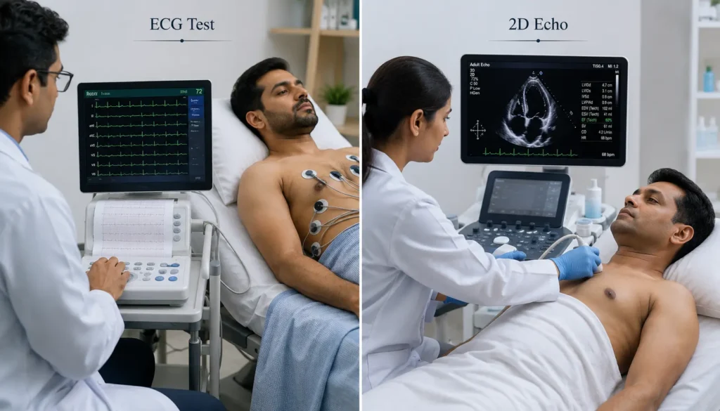
An ECG (Electrocardiogram) records the electrical activity of the heart and helps detect rhythm problems, irregular heartbeats, and early signs of a heart attack. It is a quick, painless, and widely available test.
A 2D Echo (Echocardiogram) uses ultrasound technology to show the heart’s structure, pumping function, valve health, and Ejection Fraction (EF) levels. It gives a deeper visual picture of how the heart is actually working.
Both tests serve different purposes—ECG is faster and ideal for detecting electrical issues, while 2D Echo provides a detailed structural view. Doctors often recommend both together for a complete cardiac evaluation.
Can EF Improve Again?
Yes—and this is genuinely encouraging news.
In many patients with mildly reduced heart function, EF can improve with proper treatment and healthy lifestyle changes. Even when EF 60% in 2D Echo is not present initially, the heart often has the ability to recover over time.
According to Braunwald’s Heart Disease, one of the most trusted cardiology textbooks, the heart can improve significantly when given the right support and treatment.
Controlling normal blood pressure consistently reduces the heart’s workload and allows gradual recovery. Managing cholesterol prevents further coronary artery damage. Regular moderate exercise—as advised by your cardiologist—strengthens the heart muscle over time.
Medicines like beta-blockers and ACE inhibitors can improve heart function over time. Research supports this approach, especially in patients with reduced EF before reaching normal EF 60% in 2D Echo levels.
Quitting smoking, reducing salt, maintaining a healthy weight, and attending regular follow-up appointments all contribute. Do not underestimate what consistent lifestyle care can do for your heart.
When Should You See a Cardiologist?

Even if your EF 60% in 2D Echo report is completely normal today, certain symptoms should never be ignored.
Chest pain or pressure during physical activity—even mild—could indicate Angina and needs evaluation. Sudden severe breathlessness at rest or while lying down is an emergency. Unexplained fainting or near-fainting needs urgent attention.
Swelling in both legs that appears suddenly and worsens over days is a red flag. If a previous report showed normal EF and your new report shows any drop — even to 50% — discuss it with your cardiologist promptly.
Do not wait for symptoms to become unbearable. Early detection is always better than late intervention.
Frequently Asked Questions (FAQs)
Q1. Is EF 60% in 2D Echo report considered normal?
Yes. Completely normal. The American Heart Association defines normal EF as 55% to 70%. EF 60% sits right at the healthy midpoint.
Q2. Is EF 60 normal for all age groups?
Yes. According to research published in NIH and ACC guidelines, EF 60% is normal across all adult age groups, both men and women.
Q3. What EF percentage is dangerous?
EF below 40% is clinically significant. Below 30% is severely reduced and requires urgent medical management.
Q4. Can EF 60% drop over time?
Yes, if conditions like high blood pressure, high cholesterol, or heart disease are not managed. Regular checkups help detect any change early.
Q5. Can I have normal EF in 2D Echo and still have heart problems?
Yes. Diastolic dysfunction, valve disease, or coronary artery narrowing can cause symptoms even when EF is normal. Always review the full report with your cardiologist.
Q6. Do I need treatment if my EF is 60%?
No specific treatment is needed for EF 60% itself. Your doctor will manage any underlying conditions found elsewhere in the report.
Q7. How often should I repeat a 2D Echo?
If your current report is normal, your doctor will advise based on your symptoms and risk factors. Patients with known heart conditions typically repeat every 6 to 12 months.
Final Thoughts
EF 60% in 2D Echo report is not a warning. It is not dangerous. It is one of the most reassuring numbers you can see on a cardiac report.
The goal of a 2D Echo is not to find a perfect score—it is to understand how your heart is truly functioning. And EF 60% tells us clearly—your heart is pumping well, your left ventricle is strong, and your cardiac function is within the healthy normal range.
If other findings in your report concern you, always discuss them with your cardiologist. One number never tells the whole story. But EF 60 in 2D Echo—that number tells a good story about your heart.
Medical Disclaimer:
This article is written for general educational awareness only. It does not constitute medical advice, diagnosis, or treatment. Always consult a qualified cardiologist to interpret your 2D Echo report in the context of your individual clinical history.
About the Author
Iraphan Khan, BSN | D.Pharm | CMLT, is a Healthcare SEO Strategist and Medical Content Writer at RealMedVision, creating clinically accurate content optimized for Google and AI search.
Medically Reviewed By
Dr Praveen Verma, MBBS, MD — Diagnostic & Pathology
Dr Himanshu Morya MBBS — Clinical Accuracy & Patient Safety
Kalpna Singh Shekhawat BSN NP — Patient Care & Practical Accuracy
References:
- American Heart Association — Ejection Fraction Heart Failure Measurement
- American College of Cardiology — Heart Failure Classification by EF
- Mayo Clinic — Ejection Fraction: What Does It Mean?
- NIH StatPearls — Left Ventricular Ejection Fraction
- Cleveland Clinic — Ejection Fraction Guide
- American Society of Echocardiography — Chamber Quantification Guidelines 2015
- British Heart Foundation — How to Read Your Echocardiogram Report
- Braunwald’s Heart Disease — A Textbook of Cardiovascular Medicine
- World Health Organization — Cardiovascular Disease Prevention Guidelines
- AIIMS New Delhi — Cardiology Department Clinical Protocols
- PubMed — Normal LVEF Range Meta-Analysis 2025 (PMC12828944)
- European Association of Cardiovascular Imaging — Echocardiography Recommendations